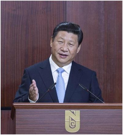
Water, forests, mountains, grasslands, wasteland, beaches, oceans, mineral resources, agricultural land, construction land, etc. all belong to natural resources. Economic theory refers to them as land. The "Decision" of the Third Plenary Session of the 18th CPC Central Committee put forward: "Improve the national natural resource asset management system, and uniformly exercise the responsibilities of the owners of all natural resource assets of the whole people. Improve the natural resources supervision system and uniformly exercise all the duties of controlling the use of land and space. " This is a major adjustment of the Party Central Committee’s thinking on state governance, which was specifically explained by the General Secretary of the Supreme Leader at the Third Plenary Session of the 18th CPC Central Committee. He pointed out: "The plenary session decided to put forward the requirements for improving the national natural resource asset management system. The general idea is to implement the ownership of natural resource assets owned by the whole people and establish a system to uniformly exercise the duties of the owners of natural resource assets owned by the whole people in accordance with the principle of separating owners from managers and managing one thing by one department. " He also pointed out: "The state’s exercise of ownership and management of natural resource assets owned by the whole people is different from the state’s exercise of supervision over natural resources within the territory. The former is the right in the sense of the owner, while the latter is the power in the sense of the manager. This requires improving the natural resources supervision system, uniformly exercising all the duties of controlling the use of land and space, so that the owners of state-owned natural resources assets and the national natural resources managers are independent, mutually cooperative and mutually supervised. "
First, the key to the reform of natural resources management is to solve the problem of no distinction between ownership and management.
In China, some natural resources are owned by the whole people (that is, state-owned), and some are owned by rural collectives. How to use the natural resources owned by the collective is the object of government supervision, and the owners and managers are separated. However, it is not clearly defined who will represent the asset rights and interests of natural resources owned by the whole people. In practice, it is often the administrative departments of governments at all levels who exercise the functions of owners’ representatives, so-called "referees" and "athletes" in one. Many outstanding problems in China’s natural resources management are caused by this.
1. The goal of government administrative supervision is difficult to achieve.
For example, in order to protect agriculture, especially grain production, the government has made strict protection of cultivated land a basic national policy, set clear protection objectives, and put forward the policy of intensive use of existing land. But for a long time, almost without exception, the development of cities all over the country has expanded to Zhang Zhilu, occupying a large number of high-quality cultivated land, and the urban stock of construction land is extensive, inefficient and wasteful. The fundamental reason is that the state does not clearly define the representative of state-owned land ownership, and the specific institutional arrangements for land management actually regard the land management department of the government as the representative of state-owned land ownership and allow it to operate land by means of transfer and mortgage. In the case that the tax-sharing reform is not thorough enough and the government still undertakes the function of urban construction, the income from government land operation has become the main source of urban construction funds. The dynamic mechanism formed by this institutional arrangement enables local governments to meet the needs of urban construction only by constantly occupying cultivated land and constantly operating land.
2. It is easy to infringe upon the interests of ordinary people.
Still taking land management as an example, because the government’s land management has become the main source of funds for urban construction, in order to ensure the government’s income from land management, rural areas and farmers have become the first to suffer losses. On the one hand, the relevant system stipulates that farmers can’t share the income from land appreciation by paying land acquisition compensation at the price of agricultural land; On the other hand, it is stipulated that all land for basic and public welfare construction and commercial projects will be expropriated by the government, and collective construction land will not be allowed to enter the market, depriving farmers of their right to participate in the process of industrialization and urbanization independently and equally with land assets. The essence of these systems and regulations is to use the power of managers to seek the interests of owners, and they are still taking the road of rural support for urban development, which is bound to be strongly resisted by farmers and intensify social contradictions. The vast number of urban residents, especially the working class, are also those who suffer the loss of the interests of the government in operating land. Basic, public welfare land, the government is a net investment; Industrial land, in order to attract investment, is generally provided at low prices everywhere, and some can’t even recover the cost; The "bidding, auction and hanging" of business land is the main source of land finance. The government should use this income to balance the deficit between basic public welfare land and industrial land, and accumulate as much construction funds as possible. Due to the design of the current housing system, there is no separate supply channel for ordinary self-occupied demand, which forces the self-occupied demand to squeeze into the supply channel of developers and compete with investment and speculative demand. In this case, the "bidding, auction and hanging" of land keeps pushing up land prices and housing prices.It has greatly exceeded the purchasing power of self-occupation demand, causing serious livelihood problems.
3. Accumulate social and financial risks.
Land transfer fee is the price of land use right for several years, in fact, the government collects land rent from enterprises for several years at one time. The land rent is the deduction of the profits of the enterprise in that year, which belongs to the category of social primary distribution. For enterprises, a centralized payment of land rent for several years means an advance of future profits, which belongs to debt management. In real life, many enterprises rely on bank loans to pay land transfer fees, and the nature of liabilities is clear at a glance. Even if it is paid with its own funds, it is still a liability in essence. If the enterprise operates well and has stable profits, it can gradually pay off this debt; If the business is not good or goes bankrupt, the debt cannot be paid off; If the reproduction of enterprises is interrupted, it will eventually turn into bad debts of banks and become a problem of the whole society. According to the relevant regulations, enterprises can transfer the remaining land use rights, so as to pay off their debts and even make profits from them. However, this is only the transfer of liabilities between enterprises, and the scale of liabilities may be enlarged.
From the perspective of the whole society, for every income obtained by the government from land transfer, there is a debt of an enterprise or several individuals corresponding to it. The so-called land finance, in essence, is a development mode that relies on the future income of overdraft society to seek immediate development. The image of the statement is "spending money on food."
In recent years, it has become more and more popular for the government to mortgage or pledge land to financial institutions. This so-called land finance is the direct debt operation of the government, which is gradually repaid with future income. At present, the scale of land finance has far exceeded that of land finance, and most of the so-called "local debt" belongs to land finance. In some places, the debts of one government may not be repaid by the next or even the next governments. The solvency of some local governments is seriously insufficient, so they delay by borrowing new debts to pay off old debts. Different from normal government debt, land finance is a "big move" of bank funds, which squeezes the private financing space and is not conducive to invigorating the economy.
Two, the implementation of natural resources management reform ideas, we need to eliminate some doubts.
The "Decision" of the Third Plenary Session of the 18th CPC Central Committee clarified the reform idea of separating the owner’s rights from the administrative supervision power. To carry out this reform idea, it is necessary to change the current situation that the owner’s representative function of natural resources owned by the whole people, such as land, forests, minerals, waters, oceans and beaches, is mixed with the government’s administrative supervision function of natural resources, separate the former from the relevant administrative power, and set up specialized agencies to exercise the owner’s rights of natural resources owned by the whole people on behalf of the state. The owner’s rights of the country, like the legal property rights of other market subjects (collectives, enterprises and individuals), are equally protected by the relevant administrative powers of the government. At the same time, the government supervises natural resources, whether owned by the whole people or collectively, according to unified rules.
The central government’s reform idea is strategically located and captures the core and key of the problem. However, there are still some people in the society, even within the party and among the ranks of cadres who have doubts about this. Some people worry that the government will no longer operate land, which will affect the local economic development and think that this is a "self-destruction of the Great Wall"; Some people worry that fair compensation for land acquisition by the state with reference to the market price will create new "rich" groups and will not benefit all farmers; Some people worry that giving rural collective construction land equal access to the market with state-owned land will make farmers, driven by interests, disobey state planning and land use control, encourage land speculation and merger by powerful groups, and encourage rural grassroots cadres to take bribes and bend the law, run amok and so on.
Practically speaking, these fears are not entirely unreasonable, and some situations may indeed happen. At present, the central government has not made specific arrangements to implement the reform, and raising possible problems will help everyone think more thoughtfully and comprehensively, and avoid or reduce the confusion that arises in a hubbub. However, it must be pointed out that some people are actually skeptical or even opposed to the central government’s reform ideas.
They didn’t see that the Third Plenary Session of the 18th CPC Central Committee decided to put forward a comprehensive reform plan, and the problems that may arise from a single reform will be avoided or corrected through other reform measures. For example, after the separation of owners’ rights and managers’ rights, although the administrative departments of the government have no income from land management, the promotion of fiscal and taxation system reform can better ensure the financial resources needed by the government to perform its duties, and the reform of investment system can expand the sources of local construction funds, and local development will be more healthy and sustainable.
They even fail to see that the main task of the current reform is to solve the problem of the relationship between the government and the market and strengthen the regulation of the government’s behavior. The local government is a combination of "referees" and "athletes", and the power is out of control, which will inevitably encourage some cadres to develop the concept of omnipotent and domineering, and fundamentally reverse the relationship between "public servants" and "masters". Only by resolutely breaking the barriers of interests with the spirit of "a strong man breaks his wrist", as repeatedly emphasized by the central leadership, can our government not be divorced from the masses and have strong credibility. The government’s behavior is standardized, and any problems that happen to the people and ordinary people are not difficult to solve.
Third, to promote the reform of natural resources management, it is necessary to break the departmental division management system.
General Secretary Supreme Leader’s speech at the Third Plenary Session of the 18th CPC Central Committee incisively analyzed the disadvantages of the current natural resources management system. He said: "Use control and ecological restoration must follow the laws of nature. If tree growers only plant trees, control water, and protect fields, it is easy to lose sight of one thing and lose sight of another, which will eventually lead to systematic ecological damage." The general secretary’s analysis pointed out that the key of the current system is to attend to one thing and lose another, and to be constrained by each other, so as not to form a joint force. Below, try to give some specific performances:
1. It is difficult to thoroughly understand the "family background" of natural resources.
Under the system of department in charge, every department should make a national plan and conduct a national resource survey for this purpose. However, because various departments use different investigation techniques and adopt different technical standards, the results of the investigation are very different and even "fight" with each other. For example, since the 1980s, the land management department has conducted the first detailed land survey for 10 years, claiming to be the first time in the history of China to find out the property of the land. According to the detailed investigation results, there were 1.951 billion mu of cultivated land in China in 1996. Later, the land management department proposed that the "red line" of the national cultivated land should not be less than 1.8 billion mu. However, on the eve of the 17th National Congress of the Communist Party of China in 2007, the forestry management department announced that from 1999 to 2007, the country’s "returning farmland to forests" was 365 million mu. According to this calculation, 1.951 billion mu is reduced by 365 million mu, leaving less than 1.6 billion mu. How can the "red line" be guaranteed? A number of forestry departments denied the survey results of the land department that lasted for 10 years! Since the state pays a fixed financial subsidy for every mu of "returning farmland to forests", this figure of the forestry department is well-founded. As a result, from the leadership of the government to all walks of life concerned, we are very confused, and it seems that the family background of our land is still unclear, and a second investigation is needed. Now the results of the second land survey have been published. With this latest data, I am afraid that some data of other departments have become a confused account again.
2. It is easy to produce regulatory blind spots.
For example, environmental protection in a broad sense should include the supervision of ecological environment changes caused by land use changes. However, under the departmental management system, the land management department is responsible for the management of land use, and the environmental protection department can only mainly manage pollutant discharge. The main responsibility of land management is not environmental protection, which leads to the absence of environmental protection. At one time, the land management department encouraged all localities to develop unused land such as coastal beaches and low hills and gentle slopes in order to "guarantee both development and cultivated land". I don’t know that the so-called unused land is just unused by human beings. These unused lands play an extremely important role in the ecological environment balance of the whole nature. Once human development activities break this balance, it will bring disastrous consequences to nature, including human beings themselves.
3. The supervision of departments conflicts with each other.
Because the resource data of different departments are inconsistent, the management based on the data will conflict. For example, if the land management department identifies it as wasteland or unused land, the forestry department may identify it as woodland or forested land. In this way, the land consolidation activities of the land department to develop unused land will be considered by the forestry department as illegal activities to destroy forests. So that in some places, the forest public security bureau arrested the director and staff of the land consolidation center. Although these problems are related to the quality of staff in various departments, the fundamental reason lies in the unreasonable management system. As the General Secretary of the Supreme Leader pointed out: "It is very necessary for a department to be responsible for the control of all land and space uses within the territory, and to carry out unified protection and restoration of landscapes, forests and lakes."
Four, specific ideas for the reform of natural resources management system
1 set up a department specializing in the management of state-owned natural resources assets.
There are two major difficulties that must be solved in China’s transition from a planned economy to a market economy. First, how can state-owned enterprises get rid of the government’s intervention and protection and become the market subject of equal competition with other enterprises with various ownership systems, and at the same time, the owner’s rights and interests of the country cannot be harmed; Second, how to correctly enter the market for natural resource assets owned by the whole people will not harm the interests of other market entities, but also effectively protect the sustainable utilization of resources. At present, great progress has been made in the reform of state-owned enterprises, and the direct intervention and protection of enterprises by the government has been greatly reduced. The establishment of the State-owned Assets Management Committee to exercise the rights of investors on behalf of the state and to plan and guide the development of enterprises as a whole has further rationalized the relationship between the government and enterprises. Although many problems such as industry monopoly still need to be solved, the direction of reform is clear and the path is clear. Compared with the reform of state-owned enterprises, in the management system of state-owned natural resources assets, the owner’s rights are more and more closely combined with the government’s administrative power, and the administrative power has even become a vassal of the owner’s rights.
In accordance with the spirit of the reform of the natural resource asset management system of the Third Plenary Session of the 18th CPC Central Committee, we can consider establishing a state-owned natural resource asset management department (hereinafter referred to as the state-owned department) to exercise the owner’s rights on behalf of the state on natural resource assets owned by the whole people. This kind of state-owned rights and collective-owned natural resource assets rights are in an equal position, and they must be subject to the unified registration of the government’s real estate registration department, the unified planning and space use control of the government’s natural resources supervision department. If the right to use enters the market, they must go through the registration and change registration of the right to use, and also accept the unified management of the departments of industry and commerce, taxation and CSRC.
Some natural resources assets, such as important mineral resources and water conservancy resources, are directly managed by the central government’s own departments; Some assets, such as the allocation, lease, transfer, shareholding and joint venture of the right to use state-owned land, and the paid use of general mineral resources and water resources, can be entrusted by the central government or entrusted to the local government for management. In addition, basic and public welfare construction needs to expropriate land or other natural resources assets owned by rural collectives, which can be compensated fairly by the state-owned departments in consultation with collectives and farmers with reference to market prices. The proceeds from natural resource assets owned by the whole people shall be turned over to the central finance, incorporated into the fund management, and uniformly arranged for use according to the prescribed purposes. Therefore, the state-owned system should implement a vertical leadership system.
Maintaining and increasing the value of state-owned natural resources assets is a duty of the state-owned departments, but it is not necessarily the most important duty. The most important duty should be to make rational use and practical protection of natural resources under the supervision of relevant government departments. The so-called rational utilization includes protecting people’s livelihood with natural resources owned by the whole people. For example, for affordable housing and ordinary self-occupied housing, the supply of land should be guaranteed.
2. Establish a department to supervise all natural resources within the national territory.
The first task of this newly established natural resources management department should be to work out a new land planning in conjunction with the National Development and Reform Commission, the Ministry of Housing and Urban-Rural Development, the Ministry of Agriculture, etc., including the main functional zoning (including major agricultural products producing areas, major commodity grain bases, major ecological and environmental protection areas, and major mineral resources accumulation areas), urban and rural construction and the layout of important productive forces, and major land consolidation (including land, forests, grasslands and waters) project arrangements, and so on. This plan is different from that of any department under the current management system. It is a comprehensive medium-and long-term plan covering all development and renovation activities within the national territory, and it is also the top plan of the national planning system. The current five-year plan for national economic and social development belongs to the near-medium-term plan and is the phased implementation of national land planning. With these two plans, there is no need to keep many departmental plans now. A few departmental plans that must be preserved and some important regional development plans must be guided by and connected with national land planning. Local land planning at all levels is an extension and refinement of national planning.
The next task is to implement space use control according to the national land planning, which not only makes rational use of resources, but also effectively protects resources and ecological environment. This is probably the most important daily work of this newly formed department. The so-called space use control is to realize how to use and protect every land and its other resources above and below the ground. It is necessary to draw a large-scale planning map, because the scale is too small, and the planned use can only fall into a larger area, which is difficult to fall on every piece of land and cannot be used as the basis for daily use control. Under the current system of departmental planning, only township land use plans and urban plans can be implemented on the plots, and can be transformed into plans that can implement space use control by unifying technical standards and superimposing relevant information. In addition, space use control also includes farmland protection, ecological environment protection and other important contents.
It is also an important responsibility of this department to formulate the trading rules of natural resource assets market. It is necessary to supervise the market trading activities such as the transfer, lease, shareholding, joint venture, mortgage, transfer and sublease of various natural resources assets, ensure the fairness and justice of the market, improve the national land acquisition system and protect farmers’ property rights. The basis of market supervision is the protection of property rights, and we can consider setting up a real estate registration authority in this department. Because this work is very complicated and involves not only natural resources and assets, we can also consider setting up a separate registration authority. Taxation on the income of natural resource assets, such as value-added tax in circulation and real estate tax in retention, is mainly the responsibility of finance and taxation departments, but natural resource management departments also have the responsibility of active cooperation.
(Author: Vice Chairman china land science society)